
Leaders in Breast Cancer Treatment
At Stanford Health Care, we offer you the most advanced diagnosis and treatment options for breast cancer, no matter how common or complex the cancer type.
Our compassionate, world-renowned doctors are pioneering new ways to bring you the best care for breast cancer.
- Specialized expertise in treating all types of breast cancer from early to advanced stages. Go to Conditions Treated
- Advanced treatment options in breast cancer surgery, radiation, and medical therapy with the latest techniques research has to offer. Go to Treatments
- Team-based approach to diagnosis and treatment for seamless, coordinated care with all of your breast cancer specialists under one roof. Go to Your Care Team
- Clinical trials that can offer you earlier access to new developments in breast cancer treatment. Go to Clinical Trials
- Comprehensive support services to help you and your family focus on health and healing. Go to Supportive Services
- Easy access to imaging services, outpatient radiation and infusion therapy, and other services in multiple locations. Go to Connecting to Care


Conditions Treated
Every member of our expert team has subspecialty training in breast cancer diagnosis and treatment to bring you the expertise you need and care you can trust.
We are devoted to offering you the most effective care based on the latest scientific data. Our robust research program and clinical trials have informed many of the leading technologies and approaches used to diagnose and treat breast cancer today.
Our breast surgeons, medical oncologists, radiation oncologists, geneticists, and other specialists work side by side every day. We bring together the right team of experts to understand and treat your unique needs.
Our areas of expertise in diagnosing and treating breast cancer include:
- Early-stage invasive breast cancer
- Advanced or metastatic invasive breast cancer
- Noninvasive (in situ) breast cancer
- Recurrent breast cancer
- Treatment refractory breast cancer
- Hereditary breast cancer and high-risk counseling
- Evaluation and work-up of suspected breast cancer
- Inflammatory breast cancer
- Breast cancer during pregnancy
- Breast cancer in young women
- Male breast cancer
Our doctors help define the national protocols for breast cancer diagnosis and treatment through their active role in the National Comprehensive Cancer Network Guideline Panels.
GENETIC HIGH-RISK PATIENTS
We have one of the most established Cancer Genetics programs in the country. If you have a family history of breast cancer, we can help you and your family members understand your risk and provide guidance on how to proactively manage it.
Our pioneering work in cancer began in 1956, when we were the first in the Western Hemisphere to use a linear accelerator for cancer treatment. We have been pushing the possibilities ever since and driving innovation to provide better breast cancer care.
INNOVATION HIGHLIGHTS
- We were the first to use gene expression profiling to identify molecular subtypes of breast cancer. This groundbreaking work has had a major impact on our understanding of breast cancer biology.
- We developed some of the first protocols for breast MRI imaging and continue to lead developments in breast imaging for diagnosis and treatment planning.
- Our intraoperative radiation therapy (IORT) program, combining radiation and surgery in one procedure, was one of the first in the nation. We continue to provide the most advanced IORT services today.
We emphasize personalized care plans designed to meet your physical, emotional, and daily living needs. Our breast imaging specialists take special measures to study your unique anatomy while working hand-in-hand with specially trained breast pathologists to provide you an accurate diagnosis. Our doctors then work closely with you to develop the best treatment plan for you. Whether you are seeking a diagnosis, searching for new hope, or proactively planning ahead, we are here to help set you on the right care path.
We offer several treatments for any type of breast cancer, including surgery, radiotherapy, and medical therapy. Because our program uses a multispecialty approach, we can easily combine input from various doctors and researchers in planning your care.
Breast Cancer Surgery involves removing a tumor and surrounding tissue. Our breast cancer surgeons are experts in breast conservation, mastectomy, and breast reconstruction.
Cancer medications target cancer through medications taken orally, intravenously, or by injection. Stanford Health Care is leading the way in discovering some of the most advanced medications. Medical oncology therapies include:
Advanced radiation therapy techniques allow us to use radiation to destroy cancer cells and slow the growth of tumors, while protecting healthy nearby tissue.
Cancer tumors have unique genes or proteins called biomarkers. By identifying these biomarkers, doctors can choose a treatment designed for that cancer. Some biomarkers can also help your care team monitor the tumor during and after treatment. Our experienced pathologists perform biomarker testing and work with your doctor to develop your treatment plan.
Your wellness plays a role in the success of your treatment. We offer our Circle of Care to assist you and your family throughout your cancer journey. Whether its spiritual counseling, a new wig, or help with your finances, we’re here for you whenever you need us.
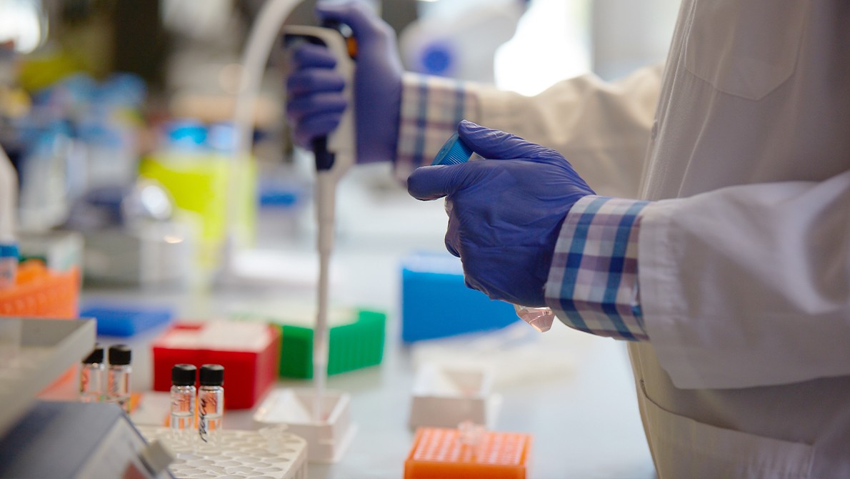
Innovation Through Clinical Research
Some of Silicon Valley’s brightest scientific minds are at the Stanford Cancer Institute and the Stanford Medicine Cancer Center. Medical discoveries and clinical advances happen here. Our physicians and scientists work every day to improve cancer prevention, detection, and care. We offer advanced diagnostics, the latest treatment technologies and techniques, and pioneering therapies in development through clinical trials. Let us put innovation to work for you.
Clinical Trials

Clinical trials are research studies that evaluate a new medical approach, device, drug, or other treatment. As a Stanford Health Care patient, you may have access to the latest, advanced clinical trials.
Open trials refer to studies currently recruiting participants or that may recruit participants in the near future. Closed trials are not currently enrolling, but similar studies may open in the future.
To learn more about the clinical trials we offer, visit: Stanford Cancer Institute Clinical Trials.
We know cancer is more than a condition—it's one of life's greatest disruptors. We offer you and your caregivers the support you need at every step of the way, including a team that actively listens, develops a plan for your unique situation, and stays with you from diagnosis to treatment and beyond.

Each person's breast cancer experience is unique, which is why we provide customized care for you. Our doctors, nurses, technicians, and staff are here to support you throughout your journey with treatment plans tailored to fit your individual needs. At weekly review meetings, experts from multiple disciplines review cases that require more complex recommendations.
Your Doctors
Medical Oncologist
Medical oncologists have specialized training in diagnosing and treating breast cancer using medications, including chemotherapy, immunotherapy, and targeted therapy.
View All {0} Medical Oncologists »Radiation Oncologist
Radiation oncologists have specialized training in using high-energy radiation to destroy cancer or prevent its spread while protecting healthy tissue.
View All {0} Radiation Oncologists »Breast Reconstructive Surgeon
Breast Reconstructive Surgeons have specialty training in rebuilding and reshaping breasts after cancer surgery to remove tumors. Many breast reconstructive surgeons are plastic surgeons and use synthetic implants or tissue from elsewhere in your body to reconstruct your breast.
View All {0} Breast Reconstructive Surgeons »Breast Surgeon
These surgeons specialize in treating cancer through traditional (open) and minimally invasive surgery. Surgical oncologists perform biopsies (taking tiny tissue samples) to test for cancer. They also surgically remove tumors, some surrounding breast tissue, and lymph nodes to evaluate them for the presence of cancer.
View All {0} Breast Surgeons »Genetic Counselors
Some cancers have a genetic component. Our counselors from the Cancer Genetics Program can guide you through decision-making about genetic testing for you and your family.
Advanced Practice Providers (APPs)
Our skilled nurse practitioners specialize in diagnosing and treating breast cancer. They see patients independently and occasionally alongside your doctor. APPs can give you a thorough exam, write prescriptions, and help prevent or treat any issues. Our APPs meet weekly to discuss patient needs.
View All {0} Advanced Practice Provider Doctors »Extended Care Team
Radiologist
A radiologist who specializes in breast cancer has additional training in imaging technologies, including X-ray, ultrasound, CT, and MRI, to detect and diagnose breast cancer. These doctors interpret imaging results and take biopsies, as needed, to help confirm a diagnosis.
View All {0} Radiologists »Pathologist
These doctors have special training in detecting and diagnosing cancer by examining tissue samples, taken during a biopsy under a microscope. Pathologists work closely with oncologists to determine the type and stage of breast cancer you have.
View All {0} Pathologists »Nurses and Nurse Coordinators (RNs)
Nurses and nurse coordinators are registered nurses who coordinate your care with your breast cancer team. They guide you from your first contact through follow-up care and help you find counseling, financial, and other support services.
Care Coordinators
Care coordinators provide you with information and assistance before and during your appointment.
- Medical Assistants: Medical assistants work with our team to help provide care. They may prepare you for an examination, assist your doctor, or take your vital signs before your appointment.
- Patient Care Coordinators: Our patient care coordinators help you with scheduling appointments and accessing your lab results. They are your first line of contact before you see your provider and guide you during your breast cancer care.
- Patient Access Representatives: Patient access representatives can answer all your questions about health insurance coverage, help you apply for health insurance, and refer you to our financial counselors.
Research Coordinators
Doctors at Stanford Medicine Cancer Center participate in research efforts to advance the understanding of breast cancer and its treatment. Research coordinators help screen candidates for possible participation in clinical research trials.
Cancer Care Services
Your cancer care includes services that focus on easing the effects that cancer and its treatment may have on you. Our support programs are available to you throughout your diagnosis, treatment, and follow-up care. Contact our Cancer Care Services for your personal support plan.
We make access to care as simple as possible. We anticipate what you need and provide support when you need it. Our network of locations puts our services within your reach. User-friendly digital health tools help you stay connected with your care team. We accept most insurance plans and offer discounted transportation, short-stay options, and international travel and translation services. We help make sense of the details, so you can make decisions that are right for you.
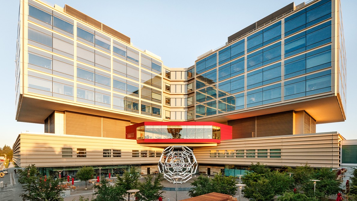
Accessing care for cancer treatment should be easy and convenient. That’s why we offer services for breast cancer at multiple locations throughout the Bay Area.
For Referring Physicians
PHYSICIAN HELPLINE
Fax: 650-320-9443
Monday–Friday, 8 a.m.–5 p.m.
Stanford Health Care provides comprehensive services to refer and track patients, as well as the latest information and news for physicians and office staff. For help with all referral needs and questions, visit Referral Information.
You may also submit a web referral or complete a referral form and fax it to 650-320-9443 or email the Referral Center at ReferralCenter@stanfordhealthcare.org.
HOW TO REFER
Email or fax a cancer referral form with supporting documentation to ReferralCenter@stanfordhealthcare.org or 650-320-9443.
To request an appointment with one of our breast cancer specialists, call 650-498-6000.