
- Specialized expertise in diagnosing and treating rare cancers of the bones and soft tissues such as muscle, fat, cartilage, and other connective tissues. Go to Conditions Treated
- Advanced treatment options that include a wide range of surgical approaches, cancer medications, and radiation therapy, often used in combination. Go to Treatments
- Team-based approach from an experienced team of doctors, surgeons, and other cancer specialists with additional years of advanced training in all types of sarcomas. Go to Your Care Team
- Clinical trials that give eligible patients access to new sarcoma therapies and are reshaping the landscape of sarcoma treatment. Go to Clinical Trials
- Comprehensive support services to help you and your family focus on health and healing. Go to Supportive Services
- Ease of access to your care team through our multidisciplinary clinics where you can see a number of specialists, often in the same visit. Go to Connecting to Care


Conditions Treated
Sarcomas are cancers that start in bones and soft tissues, such as fat, muscle, blood vessels, cartilage, and other connective or supportive tissue. These cancers can develop anywhere in the body, most often in the legs, arms, and trunk.
Because sarcomas are rare, not all doctors have experience treating them, and their symptoms can be similar to those of injuries or the aging process. It's important to get care from doctors with expertise in recognizing the signs of these rare cancers for an accurate diagnosis and effective treatment. At Stanford Health Care, you’ll find specialists with in-depth knowledge and experience in diagnosing and treating all types of sarcomas.
Doctors have identified more than 50 types of soft tissue sarcomas. Some of the more common types are:
- Angiosarcoma in the lining of blood vessels or lymph vessels
- Undifferentiated pleomorphic sarcoma in the arms, legs, or inside back of the abdomen
- Liposarcoma in fat cells in the thighs, behind the knee, or inside back of the abdomen
- Leiomyosarcoma in smooth muscle tissue in the stomach, small intestines, bladder, or uterus
Bone sarcomas
Sarcomas can occur in people of any age, but bone sarcomas develop more often in children than in adults. The most common types of bone sarcomas we treat include:
- Osteosarcoma
- Chondrosarcoma
- Ewing’s sarcoma
Sarcoma Treatments
At Stanford Health Care, you receive personalized care from a multispecialty team with deep expertise in sarcoma in all its forms. From diagnosis through treatment and follow-up, our compassionate, dedicated staff place you at the center of care.
INNOVATION HIGHLIGHTS
- Our sarcoma surgeons perform innovative surgical techniques to treat people with even the most difficult, complex sarcomas.
- Through a grant from the National Institutes of Health, we are investigating the use of magnetic resonance–guided high-intensity focused ultrasound (MRgHIFU) to treat soft tissue sarcomas.
- Our medical oncologists are studying new targeted therapy medications to treat various types of sarcoma.
Your personalized treatment plan starts with a detailed diagnosis. Your doctor carefully evaluates your symptoms and medical history and orders one or more tests. If you have previous test results, we review them and may recommend additional testing.
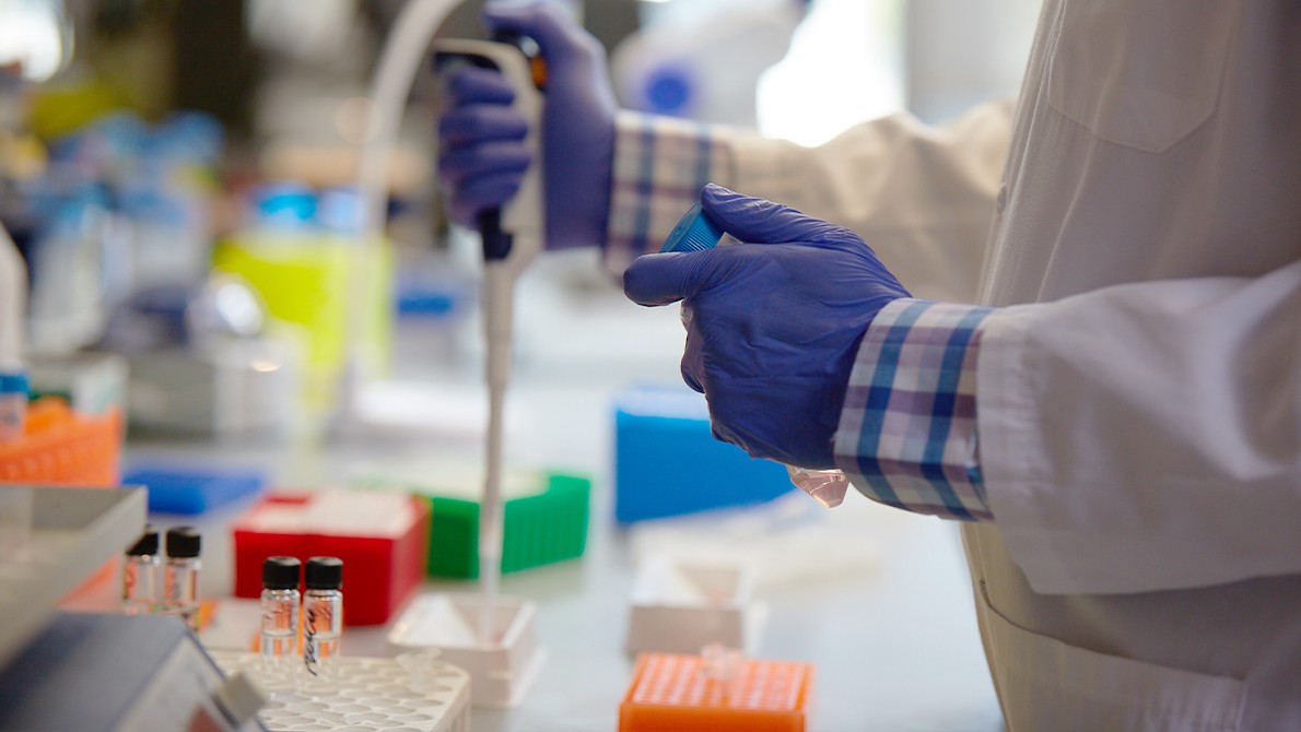
Stanford Health Care has a wide range of diagnostic tools available. Our specialists in medical imaging (radiology) and tissue analysis (pathology) work closely with your doctor to accurately diagnose your condition so you can start treatment right away.
The tests we use to diagnose sarcoma include:
- Blood tests: Sarcomas can cause changes that appear in your blood. Lab tests can help your doctor evaluate the cancer and your overall health.
- Advanced imaging: We use imaging studies such as X-rays, ultrasounds, and PET, CT, and MRI scans to evaluate the size and location of sarcomas. Your doctor may also recommend a bone scan to find out if the cancer has spread beyond the original tumor location.
- Biopsy: If your exam or imaging identifies a possible sarcoma, you will need a biopsy. During this procedure, your doctor collects a small amount of bone or soft tissue. A pathologist looks at the tissue under a microscope for cancerous cells.
- Genetic testing: Some types of sarcoma are inherited. If you have a sarcoma, your doctor may recommend genetic testing. These tests can show whether you have inherited gene changes (mutations) that might increase your family’s risk of developing sarcoma.
Your treatment plan is a care path customized to your unique condition and consistent with your treatment goals. Our cancer specialists meet weekly to review new and challenging cases to recommend treatment options. These meetings ensure multispecialty collaboration around your case and a treatment approach that meets your needs.
Surgery is often the first treatment for many types of sarcoma. Our goal is to remove as much of the tumor as possible while preserving healthy tissue.
In the past, doctors often had to amputate a limb to treat large sarcomas. Today, new techniques allow surgeons to spare the limb in most cases.
We frequently combine surgery with other treatments, such as radiation therapy, chemotherapy, and targeted therapy.
We use many types of oral or intravenous (IV) medications to destroy cancer cells or slow their growth. Your treatment options depend on the type of sarcoma and may include one or a combination of medications, including:
Stanford Health Care researchers are actively developing new forms of targeted therapy for sarcoma. Targeted therapy agents recognize specific molecules on cancer cells and attack those cells but leave noncancerous cells alone. Talk to your doctor to find out if you are eligible to participate in one of our targeted therapy clinical research trials.
Radiation therapy uses high-energy X-rays to damage or destroy cancer cells. Radiation therapy can come from an inside (internal) or outside (external) source.
Internal radiation therapy (brachytherapy) uses small radioactive beads that doctors surgically place inside or next to the tumor.
With external radiation therapy, our radiation oncologists deliver controlled radiation beams to tumors while avoiding surrounding healthy tissues. We use several types of external radiation therapy to treat sarcomas, including:
- 3D conformal radiation therapy (3D-CRT)
- Intensity-modulated radiation therapy (IMRT)
- Intraoperative radiation therapy (IORT)
- Stereotactic ablative radiotherapy (SABR/SBRT)
Our interventional radiologists deliver cancer treatment directly to the tumor using minimally invasive procedures. The types of interventional radiology treatments we use for sarcomas include:
Some cancers have a distinct genetic profile. Our experienced cancer geneticists can identify a tumor’s unique biomarkers, including genes, proteins, and other substances. Biomarkers help doctors identify tumors and match a treatment that will be most effective.
At Stanford Health Care, you receive support beyond your medical needs. We’re here to help you and your loved ones emotionally, spiritually, and socially. Our Circle of Care services are designed to ease your stress so you can focus on healing.
Innovation Through Clinical Research
Some of Silicon Valley’s brightest scientific minds are at the Stanford Cancer Institute and the Stanford Medicine Cancer Center. Medical discoveries and clinical advances happen here. Our physicians and scientists work every day to improve cancer prevention, detection, and care. We offer advanced diagnostics, the latest treatment technologies and techniques, and pioneering therapies in development through clinical trials. Let us put innovation to work for you.
Clinical Trials

Clinical trials are research studies that evaluate a new medical approach, device, drug, or other treatment. As a Stanford Health Care patient, you may have access to the latest, advanced clinical trials.
Open trials refer to studies currently recruiting participants or that may recruit participants in the near future. Closed trials are not currently enrolling, but similar studies may open in the future.
To learn more about the clinical trials we offer, visit: Stanford Cancer Institute Clinical Trials.
We know cancer is more than a condition—it's one of life's greatest disruptors. We offer you and your caregivers the support you need at every step of the way, including a team that actively listens, develops a plan for your unique situation, and stays with you from diagnosis to treatment and beyond.
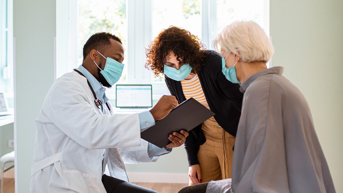
We use a team-based approach that prioritizes collaboration and ensures coordinated and personalized care. Our doctors, specialists, advanced practice providers, nurses, and staff are dedicated to your health and well-being.
Your Doctors
Medical Oncologist
Medical oncologists have specialized training in diagnosing and treating cancer using medications, including chemotherapy, immunotherapy, hormone, and targeted therapy.
View All {0} Medical Oncologists »Radiation Oncologist
Radiation oncologists have specialized training in using high-energy radiation to destroy cancer or prevent its spread while protecting healthy tissue.
View All {0} Radiation Oncologists »Hepatobiliary Surgeon
These surgeons specialize in treating cancers of the liver, bile duct, and pancreas. Cancer surgeons perform biopsies (taking tiny tissue samples) to test for cancer. They also surgically remove tumors and surrounding tissue to evaluate for the presence of cancer.
View All {0} Hepatobiliary Surgeons »Surgical Oncologist
These surgeons specialize in treating cancer through traditional (open) surgery and minimally invasive procedures. Cancer surgeons perform biopsies (taking tiny tissue samples) to test for cancer. They also surgically remove tumors and surrounding tissue to evaluate for the presence of cancer.
View All {0} Surgical Oncologists »Orthopaedic Surgical Oncologist
Orthopaedic surgical oncologists use innovative surgical techniques to treat cancer of the bone. They have expertise in limb salvage surgery, which often allows surgeons to remove tumors while preserving the arm or leg.
View All {0} Orthopaedic Surgical Oncologists »Thoracic Surgeon
These surgeons specialize in treating cancer through traditional (open) and minimally invasive surgery (video-assisted thoracic surgery and robotic-assisted thoracic surgery) to remove tumors. They are also experts in recommending other forms of cancer treatment to be used in combination with surgery or instead of surgery in cases where surgery is not appropriate.
View All {0} Thoracic Surgeons »Advanced Practice Provider
Our skilled nurse practitioners specialize in diagnosing and treating sarcoma. They see patients independently and occasionally alongside your doctor. APPs can give you a thorough exam, write prescriptions, and help prevent or treat any issues. Our APPs meet weekly to discuss patient needs.
View All 4 Advanced Practice Providers »Extended Care Team
Nurses and Nurse Coordinators (RNs)
Nurses and nurse coordinators are registered nurses who coordinate your care with your sarcoma team. They guide you from your first contact through follow-up care and help you find counseling, financial, and other support services.
Care Coordinators
Care coordinators provide you with information and assistance before and during your appointment.
- Medical Assistants: Medical assistants work with our team to help provide care. They may prepare you for an examination, assist your doctor, or take your vital signs before your appointment.
- Patient Care Coordinators: Our patient care coordinators help you with scheduling appointments and accessing your lab results. They are your first line of contact before you see your provider and guide you during your sarcoma care.
- Patient Access Representatives: Patient access representatives can answer all your questions about health insurance coverage, help you apply for health insurance, and refer you to our financial counselors.
Genetic Counselor
Some cancers have a genetic component. Our counselors from the Cancer Genetics Program can guide you through decision-making about genetic testing for you and your family.
Research Coordinators
Doctors at Stanford Medicine Cancer Center participate in research efforts to advance the understanding of sarcomas and their treatment. Research coordinators help screen candidates for possible participation in clinical research trials.
Cancer Support Services
Your cancer care includes services that focus on easing the effects that cancer and its treatment may have on you. Our support programs are available to you throughout your diagnosis, treatment, and follow-up care. Contact our Cancer Care Services for your personal support plan.
We make access to care as simple as possible. We anticipate what you need and provide support when you need it. Our network of locations puts our services within your reach. User-friendly digital health tools help you stay connected with your care team. We accept most insurance plans and offer discounted transportation, short-stay options, and international travel and translation services. We help make sense of the details, so you can make decisions that are right for you.
Accessing care for cancer treatment should be easy and convenient. That’s why we offer services for sarcoma at multiple locations throughout the Bay Area.
For Referring Physicians
PHYSICIAN HELPLINE
Fax: 650-320-9443
Monday–Friday, 8 a.m.–5 p.m.
Stanford Health Care provides comprehensive services to refer and track patients, as well as the latest information and news for physicians and office staff. For help with all referral needs and questions, visit Referral Information.
You may also submit a web referral or complete a referral form and fax it to 650-320-9443 or email the Referral Center at ReferralCenter@stanfordhealthcare.org.
HOW TO REFER
Email or fax a cancer referral form with supporting documentation to ReferralCenter@stanfordhealthcare.org or 650-320-9443.